Category:Plasma Scientific Journal
Contents
- 1 Articles published in 2018
- 1.1 October
- 1.2 September
- 1.3 August
- 1.4 July
- 1.4.1 Making Plasmatic Fields Visible and Measurable
- 1.4.1.1 OBJECTIVE
- 1.4.1.2 METHOD
- 1.4.1.3 HISTORY
- 1.4.1.4 PREPARATION
- 1.4.1.5 SETUP
- 1.4.1.6 METHODS OF OBSERVATION
- 1.4.1.7 BASIC CLASSIFICATION
- 1.4.1.8 SHORT EXAMINATION WITH DETAILS OF THE IMAGES
- 1.4.1.9 COMPARING THE RESULTS
- 1.4.1.10 SPECIAL OBSERVATIONS
- 1.4.1.11 A SURPRISE
- 1.4.1.12 SPECIAL OBSERVATIONS
- 1.4.1.13 CONCLUSION
- 1.4.1.14 REFERENCE
- 1.4.1 Making Plasmatic Fields Visible and Measurable
Articles published in 2018
October
.
September
Fukushima Decontamination Solution as a Gift for Humanity
Introduction
The origin of this paper is based on the first video, which were released by the Keshe Foundation about how simply the radiation and the radioactive contamination in the farms and the environment around the disaster zone of the Fukushima area was shown to be decontaminated, by the use of new materials developed by the M. T. Keshe.
This, after the tsunami of the 2011, after three years can be resolved, simply by the use of basic materials, farmers on their own and by the community can clean up the environment, rather than with the help of the governmental organizations.
The video is available at: https://www.youtube.com/watch?v=4f02CcnHjSk
Experiments
One of our Knowledge seekers performed a series of experiments with decontamination of radioactive materials using procedures developed by Keshe Foundation and materials prepared by us in its Spaceship Institute in Italy. We were connected with and helping her during this experiments over Skype calls. Gamma ray detectors were used. These shortcuts are used for physical quantities:
• A [Bq] … radioactivity, counts of decayed particles per second
• S [Bq / kg] … specific radioactivity, radioactivity per unit weight
Measured values in ( ) brackets are under measurable threshold of the detectors.
Numbers in [ ] brackets refer to attached measurements.
Experiment in food assurance company in Fukushima Region
Sample 1, March 29, 2014
Contaminated radioactive soil from a rice field.
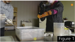
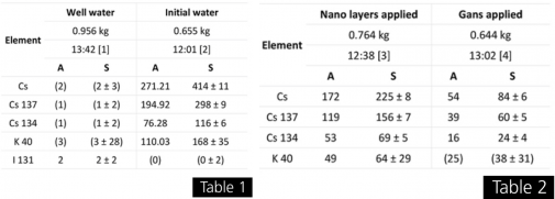
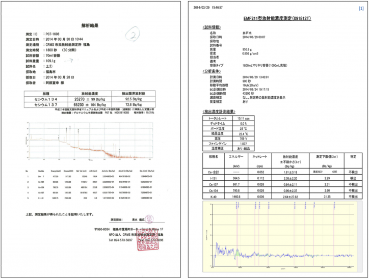
The soil in a box was soaked with well water (Table 1) and few drops of detergent (to allow better release of radioactive elements into the water) and stirred well (Figure 1).
The water from the top of the box was poured into a test container so that most of the soil sediments were kept in the box.
Half of the water from the test container was poured into another test container to remove more soil sediments to get initial water (Table 1).
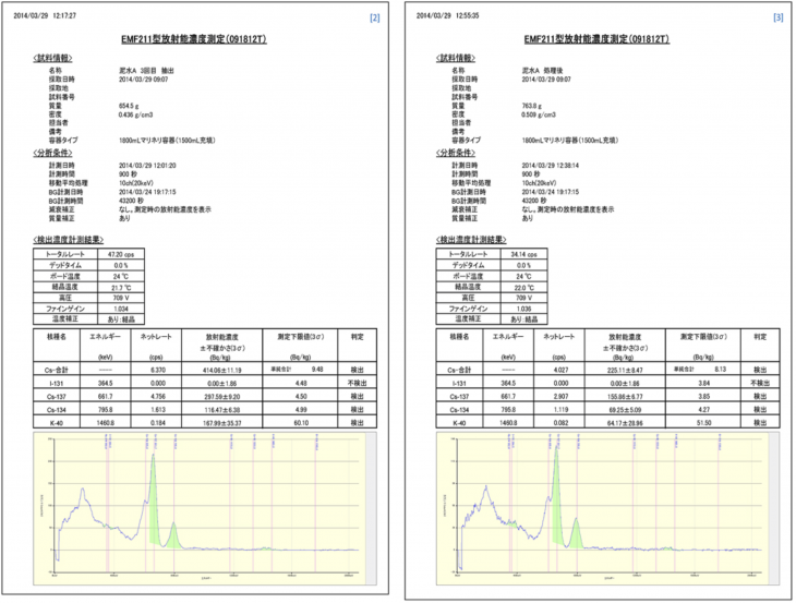
Part of initial water was treated with cylinder filter with nano layered Copper wires (Table 2) while well water was added to make the filtering simpler (Figure 2-4).
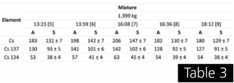
The rest of initial water was treated with Copper compounds material in GANS state. (Table 2, Figure 5). Both previous treated water samples were put together to final mixture (Table 3, Chart 1, Figure 6). Last measured decrease in radioactivity of Cs was 33% relative to initial value. Video recording of this experiment is available at: https://www.youtube.com/watch?v=7hCk4HIjzWk&index=5&list=PL2tl5CAdIAVhqirHaZWVlk_v-bBE0oFFb
Sample 2, March 31 – April 3, 2014
Contaminated radioactive water extracted from a rice field soil with the help of iron […]
Experiment in NPO in Fukushima city
Sample 1, March 30, 2014
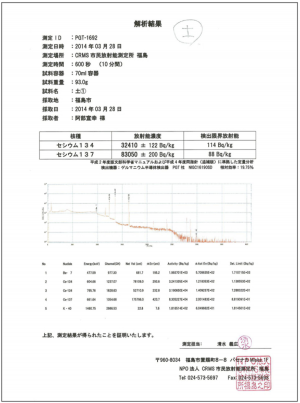
Contaminated radioactive soil from a roof gutter […]
Sample 2
Contaminated radioactive water […]
Experiments Summary
Over the measured period of time, all methods showed decreased values of radioactivity in the samples. This decrease was caused by direct transformations of the radioactive material and not by separating the radioactive material by any method of absorption out of the samples (except the cases with nanolayer filters of course). These experimental verifications of the decontamination technology have to be further developed in cooperation with people willing to take part in the global clean up. We are here to help in this process and learn more together to achieve simple real-world application of the disclosed decontamination process.
Knowledge gained
[…] The conclusion from the development of new materials and their use in the contaminated environment of Fukushima and any other reactive environment, like a nuclear waste disposal and storage is as follows:
1. All radioactive contaminated environments and materials can be made fully and permanently radiation safe.
2. The new process of decontamination can bring about new opportunities to convert the radioactive materials to useful and applicable materials, which can be used for scientific development, agricultural use, and energy production for conversion of matters.
3. These tests have shown without a shadow of a doubt that the Japanese Fukushima disaster of 2011 can be used to the advantage of the nation and not as it looks, as a disadvantage and a financial burden.
4. All underground storage facilities and nuclear waste problems of today can be solved with little effort and little cost by the use of these new materials, and there is no need for these heavy radioactive materials to be kept in storage for hundreds of years for them to be less radioactive, now one can safely use the by-products of decontamination for the production and their use in agriculture and material production rather than wasting money to store them for years on end.
5. The test shows clearly that The Fukushima plants we use for the production of plutonium. As a nuclear engineer and being involved in the promotion of peace, I see this as unacceptable and against the present agreed international laws of the proliferation of the UN charter. These tests have shown a new and brighter life for humanity and hopefully, as a race, we become stronger with the gained knowledge. […]
Plasma Applications in Agriculture
Effects of Co2, CH3 and CuO Liguid Plasmas on Early Seedling Growth of Oats and Maize
August
Gravitational-Magnetic Battery
OBJECTIVE
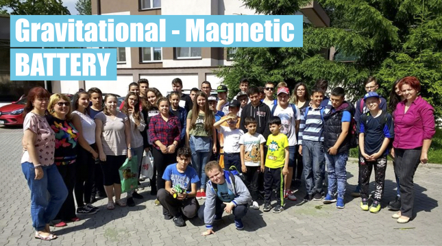
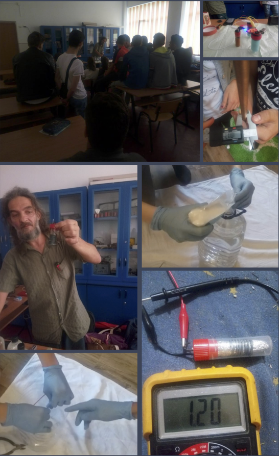
Gravitational Magnetic Battery, made by D. I., fifth grade schoolboy at I.D, Sirbu of Petrila, Hunedoara county, Romania. Presented at the Minitehnicus County Contest, organized at the Children's Club in Petrosani on 9 June 2018.
METHOD
Method: Copper wire 2,5 mm nanocoated with flame 4 times.
GANS used:
10 ml GANS mix - 40% CH3, 18% CO2, 18% ZnO, 18% CuO.
PREPARATION
Classic GANS made with led connection and nano coils covered by the flame method and 10% salts.
SETUP
We used a 9 cm nanocoated copper wire that was rolled over a paper towel that had a mix of GANS. I wrapped the paper napkin with an aluminum foil in the kitchen of 28 cm long and the width of 7, to which I bent 1 cm (from the end). Aluminum overwrap was 9 turns in anti-clockwise direction with 2 cm zinc wire springs.
APPLICATION
This battery was tested at the Minitehnicus contest in date 9 June 2018 in Petrosani city, Romania.
OBSERVATIONS
After this plasma battery was developed, a procedure to improve the result was found by replacing the paper briefcase with cotton disks used for women's cleanser.
RESULTS
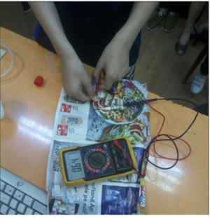
The Minitehnicus contest measured the battery with a multimeter, and the registered value was 0.64 Volti. The responsible student received the diploma of the 1st place.
REFERENCE
We mention that D. I. requested to be educated with the information from the plasma science and participated in the theoretical and practical workshops that were made by the members of Plasma Hunedoara Study Group, the Local Laboratory in Petrila, coordinated by Giani Marin Boia and Pelacaci Georgeta Emilia, who are preparing 3 high school persons for Vietnamese lab competitions, along with Physics teacher Cindea Nicoleta from Constantin Brancusi Technical College in Petrila. This project is carried out in collaboration between the "Constantin Brancusi" Technical College in Petrila, with the support of the Plasma Romania Scientific Association and the Keshe Romania Association, and National Institute for Research Development for Mining Security and Explosive Protection. Thank you, Mr. Keshe.
ALS - A Death Wish That Comes True
[…] With the release of this paper Stichting the Keshe Foundation for the first time in its history opens the doors of its research to the public for a glimpse into one of its most closely guarded research programs. […]We have developed the health section of our spaceship program because we believe the spacecraft of the future will not be able to carry all the medicines and doctors of every discipline required to cover all aspects of the health needs of people in long-haul deep space travels. In order to meet every eventual medical need there would be more medicine than food and more doctors than passengers on board these craft.
An insight into the principles by which the Keshe Foundation designs and operates its body resetting systems
In our work we consider the plasmatic structure of the elements and the interaction of the Magnetical and gravitational fields of these elements in the human body. We consider not only the physical method of connection and communication between different parts of the body, but we look at the invisible connection and interactions of magnetic fields and gravitational fields of the matters of the elements or organs in the body relative to each other. This invisible but real interaction of different parts of the body
is one reason why the world of medicine has such a hard time at present curing most ailments.
We consider the human body to be like a galaxy in the universe, with all its physical entities and the hidden and invisible interactions and connections of its magnetic and gravitational fields (Magravs) forces. In a galaxy there are visible stars and planets and at the same time these have invisible Magravs connections between all their parts and with each other. […]
New fundamental criteria used in application to health […]
From our research we can with confidence state that amyotrophic lateral sclerosis is a reversible condition, if it is discovered at the right time and handled in the right way by a competent team of physicians. In view of our development of plasma technology and its applications, amyotrophic lateral sclerosis should no longer be a death sentence handed out by doctors, who tell their patients in the same breath that there is nothing that can be done to save their life as the present world of science has no cure for this killing disease. The present medical world cannot explain the origin of amyotrophic lateral sclerosis or what triggers this process in the human body. […]
We have been striving to understand why the body puts itself through this horrendous process of amyotrophic lateral sclerosis that brings about its death. We see this as one of the illnesses most likely to occur in space when people are away from home and unknowingly can trigger such a process through depression or loneliness during long periods of being away from loved ones.
For the process to start, it requires two psychological trigger points during the life of the patient. One such event may take place in the early years or in the teenager phase and then for the illness to show itself outwardly, a second trigger point is needed. This can take place in the early twenties or in later years, causing the recurrence of the same feeling as in the first trigger point. […]
The point that has not been understood up to now is that most carbons are of diamond structure and the information received by the carbon through a given channel for a matter of a microsecond changes the characteristic of the carbon structure of the amino acid to given resistor strength level or to a given carbon structure of graphite atomic form as the carbon needs to be for a given current level of the information received from the brain for given retraction or reflex. In This process causes the potassium of the electro-motoric junction valve to be changed into insulating mode or disconnection mode, keeping the insulator crystal structure. […]
In the case of amyotrophic lateral sclerosis for example in the electro-motoric junction of the fibres, both the lines of the electric and the emotional fields operate simultaneously, with the potassium functioning to contract the fibre while the sodium process retracts or opens the fibre, which needs the lesser force. In the motoric operation, the potassium releases stronger fields and so it commands a stronger and longer contraction, as when a man needs to hold onto a bar to save himself from falling, while to open the same fingers the muscles use partly the reflex and partly a small amount of energy to allow the fibre to relax, which is done by the sodium magnetic field release. […]
Amyotrophic lateral sclerosis is not the work of one doctor; it needs a good dedicated team of physicians, psychologists, nutritionist, kinesiologist and neuro-tissue specialist to be there in every stage of development of the process and progress with the patient. Reversing amyotrophic lateral sclerosis and multiple sclerosis requires a caring and dedicated team if the case is to be successful in all aspects. […]
Wisdom gained
From our research into the cases of amyotrophic lateral sclerosis over the years and our understanding of how this process starts in the human body, we have called this illness the death wish that comes true. The affected person has unknowingly through two different but similar emotional situations in two different stages
of their lives wished they were dead, and then when their wish comes true they have to complete their unconscious desire through such a traumatically slow and painful death. The patient himself triggers the physical death-code of his own body. [..]
In all aspect of physics and the world of science man always has looked for the physical connection between the parts of the human body and has forgotten or never understood that as the human body is made of the plasma of protons and electrons and plasmas of all the atoms in the molecules of amino acid, these plasmas irradiate Magravs and receive Magravs of other cells, so they do not need a physical connection to interact
and affect each other's performance.
Amyotrophic Lateral Sclerosis
The process starts by the person wishing to be dead, and progresses through paralysis and silence to eventual death. Now with the new plasma technology developed by the Keshe Foundation and as we have seen with this case and a number of other cases around the world we can say with confidence:"We have enough knowledge now to start to reverse the process of amyotrophic lateral sclerosis in the human body psychologically, and then the body will reverse the physical damage by itself. "
On behalf of the Keshe Foundation M T Keshe
Director of Stichting the Keshe Foundation
Spaceship technology in the service of humanity
The complete published article can be found at the following link: https://usastore.keshefoundation.org/store/product/ALS/
July
Making Plasmatic Fields Visible and Measurable
Christian Böttgenbach, Student at KF SSI Education, Feb 2018
OBJECTIVE
This is the description of a method to make visible, compare and measure plasmatic (MaGrav) fields, as requested by the Keshe Foundation. It is an ongoing study, the results encourage me to share the method used and some of the results at this early stage. I want to set up a database to be able to show, determine and measure the plasmatic fields of GaNS. I use a method of creating rising pictures through a capillary-dynamic process, that has been developed by W. Hacheney.
METHOD
Any sample will release its fields with help of water into a suitable filtering paper during a capillary-dynamic process. This happens, because the fields can create micro-motion in matter state fluids, if the fluids are in an open state of matter, GaNS-like. Usually we do not see this motion created by fields, but when absorbed instead of pressed, fluids, especially waters, will freely release the fields they are carrying, in shape of micro-motion, to another medium. In this special setup this motion of the water is being braked, when it is absorbed through capillary diameters of 2 micron or less. We use metal salts to colour this otherwise invisible process. The metal salts are released, where the micro-motion slows down, giving us an exact copy of the field-induced motion of the carrier, the water.
HISTORY
Wilfried Hacheney developed and used this method to determine morphology and powers (MaGrav fields) behind the substances he has been working with as an engineer. He made about 150.000 images this way. I was taught by him how to create and analyze the resulting images. His invention corresponds to earlier developments by E. Pfeiffer, W. Kaelin, L. Kolisko and others, going back to hints by R. Steiner about 100 years ago. A more recent dissertation by Aneta Zalecka (Uni Kassel, 2006) reveals, that even the older methods of creating rising pictures are valid scientific methods, concerning comparability and evaluation of the quality of food. We met her in her lab to watch her work and discuss results.
PREPARATION
Materials
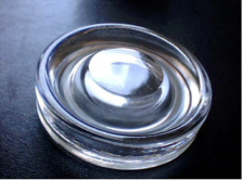
- Get Kaelin petri dishes (amorphous glass) with a rise in the middle, for the fluids to gather in a ring close to the outer rim. They can be bought at “Forschungsring Darmstadt e.V.” in Germany.
- Buy argentum nitricum (2%) and ferrum sulfuricum (2%) as well as a pipette and small bottles with pipettes for dispensation of equally sized drops. You can probably get that at your local pharmacy.
- Have gloves ready, otherwise you might create an image of your DNA. I use simple disposable latex gloves.
- Find suitable filtering paper. I use a special paper, 100 gr/m2, ca. 200 micrometer thickness, with an opening of 2 micrometer or less. My paper had been developed by Mr. Hacheney, until now I did not find anything matching its quality. I am working on that with Hahnemuehle, one of the most renowned producers of filtering and technical papers. The paper is the most important ingredient for the creation of these images. Without the right paper you might still get some pictures, but no clear, measurable shapes and relations. Blotting paper and orthochromatic paper will not work sufficiently.
- Use neutral water, it is needed as a reference and a carrier substance. All fields carried with the water will influence the images. Keep magnets, crystals and all “water-guru” stuff away from it. I use distilled water and additionally I try to bring it into the best state to be able to transfer the fields into the filtering paper. Our breath can teach us there:
The water droplets in our breath are about 2 micron in size, creating a huge surface of about 300.000 m2 per liter. This way the fields can easily be taken over by the water. I use a “levitation device” to move the water very fast ( 6x speed of sound), without pressure, into a special shape, to open it up into these small droplets. Existing fields carried by the water are being erased during that process. The water will be in the same state, have the same “inner surface” (if you add the surfaces of the micro droplets), as we have in our breath. Of course, you can do without that machinery. I just explain it to add to the knowledge and to offer an idea, what your soul might wish, when preparing the water. Cooking also helps to increase the inner surface of water and to erase some fields.
- A scanner would be handy to document the results. I scan the images with 2400 dpi, raw format and without backlight. It would be better to use a backlight to also acquire the faint shapes below the surface of the image.
No image processing at scan time recommended. Some software like “riot” to resize the images and “ImageJ” for filters, measurement and evaluation might be helpful afterwards, both are free
SETUP
- Create an environment with little disturbances from all kinds of fields and radiation, including direct light, because they might influence the process. The results are also slightly influenced by the fields of daytime, earth, phase of the moon, planets and stars. For best results, 20° Celsius and 50-60% humidity are preferable. Small deviations might result in slight changes of size and colour but you will still create a useful image.
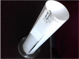
- Cut the filtering paper into sheets of 167 by 167 mm. Then make an extra cut, 25 mm from one of the borders. Bend the paper to a tube and bend the extra snippet away or cut it off, like I did on the picture. Attach a stainless paperclip to keep the paper in shape. If you use something else than a Kaelin petri dish, check the size of the paper you need beforehand.
- As it is a sensitive process and we have the same fields within ourselves, that we are creating images of, be aware of your emanations. It would be advisable to be in a balanced mood.
- Label the paper with the sample used and date of creation. Place the clean Kaelin petri dish, dispense up to 3 drops of GaNS Liquid (depending on the material to be tested) into the ring and add 4 drops of water. I use distilled and levitated water for neutral and powerful results. It might be necessary to create images of your water also, as a reference. Actually, you can examine anything this way, be it
fluids like blood (use only one drop of blood), saliva, juices from plants or hard materials or even emotions, if you add them to a fluid like water.
- Then place a suitable filtering paper, prebent to a tube, into that dish, so that it absorbs the liquid at the bottom. The orientation of the gap should be to the north.
- After about 20 minutes add 4 drops of silver nitrate solution (2%) and 3 drops of distilled water and put the paper back into the petri dish. Always check the orientation.
- After another 20 minutes add 3 drops of ferrum sulfuricum (2%) and 4 drops of water, same procedure.
- After 20 minutes again, add 2.5 ml of the water (preferably distilled and levitated) and then let it dry for about 12 hours. Remember to keep the image protected from direct light until it is dry.
- Then give it some light, diffuse daylight is fine, for development of the colours, for about one day. If you are testing other substances, it may take several days to develop them. Although sulfur stops the development of the silver, the pictures may become a little darker and loose some sharpness over time. Images may also change over time according to the state of the origin of the sample. I scan them, when they are ready.
METHODS OF OBSERVATION
The best way to observe the results would be a light box, because when observing just the surface of the paper, some faint structures will remain hidden. Placing images on a window (daylight) works very well, too. Otherwise you might want to use scans of the image, which allows to enlarge them easily. I got a special pair of compasses (Relationalzirkel) from Mr. Hacheney. He told me to pay attention to all shapes and compare their relations to each other with it. It is also possible to measure and compare everything else, the most easy thing to start with is the height of the images. All GaNS images I created until now, show a different height, depending on the GaNS used as a sample. CH3 images build up about 10% higher than CuO2 images. The most important element of all observation is unbiased perception. Do take your time to repetitively watch an image without any assumptions, until it starts to reveal its secrets. The more images you have seen, the faster and easier important correlations can be found. Gain experience, reading the pictures really is an imaginative process.
The main reason to choose this method is its exactness, you can literally see everything in these images, if you have learned to read them. I am still at the beginning but would like to mention an example of exactness, that I experienced with Mr. Hacheney: When I gave him an image of my saliva, he looked at it briefly and told me that i have got two dead teeth. I only knew of one and I could not even see specific teeth in the image then. The other day I went to a dentist and it turned out that he was right. But it was far more, what he told me about my teeth, about certain weaknesses and strengths, what will happen to them and how to bring balance and health to them. What he could read out of an image of my blood, was even more astonishing, because he could see very specific things, that where going to happen in future. This is not a miracle, because every process initially occurs in the fields, before it manifests in matter state. Knowledge seekers know that anyway.
As a child, I was in a lucky situation, like Mr. Keshe, having a father that was dealing (literally) with X-ray films. My father also sometimes had to teach physicians how to read their images and he showed some at home. Also I studied eurythmy, which helps me now to understand the characteristics and qualities of the movements of the fields, which we can see in the rising pictures. Everyone has his own background, even more so it is desirable to find some kind of classification and standardization for this process, so we can compare, determine, practice and understand, wherever we are.
BASIC CLASSIFICATION
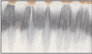
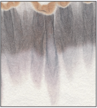
I created several series of images of CO2+ZnO, CuO2 and CH3 GaNS. I will only show one of them, all 3 images were created simultaneously. Before we can compare them, we have to find a rough classification. Enlarge the pictures and perceive them. Take your time!
The images contain several obvious elements:
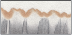
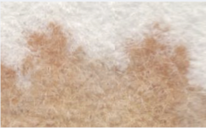
- A brownish upper horizon with a special thickness, amplitude, curvature and intensity.
A second, blurred, grayish horizon, with clear differences in thickness and intensity, interrupted by vertical cylindrical pipe shapes.
- The pipes themselves, they seem to be 3-dimensional, at least. They strongly differ in many aspects, depending on the GaNS Liquid used as sample.
A closer look will reveal many more elements. Directions, relative angels, rotations, opacity, convexity and concavity as well as repetitions, sizes and amplitude can be taken as separate elements. This work is still in its initial stage. We will continue with a simple examination of the upper horizon and the pipes.
SHORT EXAMINATION WITH DETAILS OF THE IMAGES
CO2/ZnO
Look at the brownish horizon. This sample shows an astonishing horizon there, because it has a lot of twin hills, and also alternately bigger and smaller hills. At some places, this horizon seems to fade away from below.Many of the pipes show up in pairs that seem to correlate.They regularly touch the upper horizon. Some of the darker pipes stay open at their top, where they touch the brownish horizon. Some of the single, thin and less coloured pipes seem to prick the horizon with their thin peaks.
CuO2
Here we find an irregular-shaped, rather thick horizon, with hills pointing into different directions and deep, in part narrow valleys. Below it there is a very faintly colored region.The pipes are mainly closed quite flatly at their almost colourless top, way below the brownish horizon. They are rather short and weak, unable to push through the greyish, weak belt. In many cases the colour surrounding the pipes seems to be stronger than the border of the pipes themselves. The detail images can be enlargedCH3
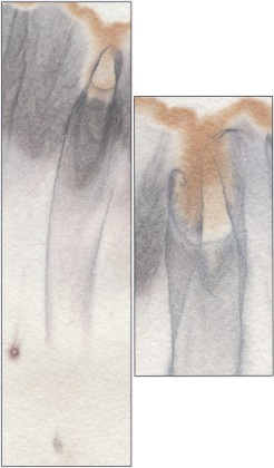
Here the brownish horizon is being superceeded by the pipes from below. It is strong but not independent, rather irregular and with a low amplitude. Observing the meandering grey lines from below shows an otherwise hidden structure that may help us to understand, how the brownish horizon is being created in general.Now these are many, big, strong and dark pipes. None of them ends at the brownish horizon or below, instead, they all stay open at their top. We can see some brownish color in the greyish layer here. Look at the structures surrounding the pipes and branching out of them. Try to imagine direction, rotation and energy of the fields at the point of creating the image.COMPARING THE RESULTS
Comparing the images will give us insight into the possibilities of rising pictures in general and it might also help to understand the characteristics of specific plasmatic fields. The first impression I want to mention here is the (at least) twofold character of the CO2/ZnO image, which can be observed in particular there. Until I have pictures of clean CO2 and ZnO apart from each other, I have a presumption: I believe, that we can see the single components of the fields of at least CO2 and ZnO there, although we learned from Mr. Keshe, that the resulting fields become a single entity. I expect, that this method allows the analysis of the combined fields and the strength, quality and even percentage of its components.
When comparing this CO2/ZnO image with the CuO2 image, we can clearly see a difference in field strength. The CuO2 image seems to be attached to the ground, probably due to more gravitational fields, compared to the environment. This would underline the importance of neutral water, that we use as a carrier. I tried to make images, where I replaced all water with the GaNS Liquid of the sample. The resulting images still allow a recognition of the kind of GaNS used, but are by far less significant. When comparing the CuO2 image with the CH3 one, we see the greatest difference between all images shown so far. The image shows a strong push upwards, or is it sucked upwards? Or even coming down from above? What do you feel about that? Some of the pipes seem to open up, becoming wider at their top. CH3 we characterize as a giver of energy and it is known to be an Magnetical GaNS. It seems that my GaNS meets this description. More tests with same kinds of GaNS from different sources have to be done.
SPECIAL OBSERVATIONS
There is a strange component at the right rim of the CH3 image, that does not seem to fit in there. Look at this strange Pipe with that little finger with fingernail in it. Something like this did not repeat in any of my images of GaNS. Still this did not happen accidentally. Look at the bottom of the picture section, two impurities can be found there. They had been on the paper before, and I do not know, what they consist of. When we really learn to read the images, we will know. I placed that here to demonstrate the exactness and beauty of the conversion of any plasmatic field into a picture.
To the right, we see a detail from the center of the CO2/ZnO image. I am stunned every time I look at that shape. Can you follow the tender movement of the semitransparent veil, do you feel the harmony of it, can you see the picture of a madonna with her child? How many dimensions does it reveal? Let it talk to your soul!
A SURPRISE
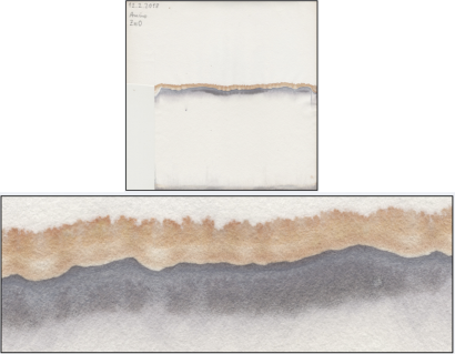
While watching the GaNS images, I had to think of amino-acids and that Mr. Keshe taught us, that they form the most beautiful star formations. This is what happened when I created an image of my ZnO amino-acid:
Above: Faint suns in the brownish horizon and below in the grey region.
Below: When really zooming into that same image, these structures appear. None of the GaNS images contain anything alike: Lots of little star-formations!
SPECIAL OBSERVATIONS
When I saw this, I knew it is time to come forward and share, what I found
CONCLUSION
Although still at the very beginning, I believe to have found a valuable method to make plasmatic fields visible, comparable and even measurable. In contrast to other methods like crystallization, nothing is forced here, the fields release themselves freely, as if they want to teach us. There is a lot of work to be done. Many images, more classification, measurements and many comparisons have to be performed to add to our knowledge. The method is flexible, low cost, significant and very powerful. It has the potential to become a standardized evaluation instrument for GaNS and plasmatic fields. I will call it “Plasma imaging”, unless otherwise advised by Keshe Foundation.
REFERENCE
All references refer to older methods of capillar dynamolysis or rising pictures, except the audio recording from W. Hacheney. The older methods are more sensitive to disturbances and give less exact but still sometimes very beautiful results.
Wilfried Hacheney, 13.3.1924 – 20.4.2010. Some of his works: Organische Physik. Aufsätze, Michaels-Verlag (Dezember 2001)
Der Weg – Der Mensch vom Geschöpf zum Schöpfer Wasser, Wesen zweier Welten. Michaels-Verlag (Dezember 2003)
Audio recording on “rising pictures” 2004/09/10, Kassel
You may also want to do a research on his patents here: https://www.dpma.de/recherche/
Friedrich Hacheney, Hyper-Wasser: Wasserenergetisierung nach Hacheney, 2014
(Wilfrieds son) Levitiertes Wasser in Forschung und Anwendung, 1994
Recent scientific works:
https://hds.hebis.de/ubks/Discover/EBSCO
lookfor=steigbild&type=allfields&service=combined&submit_button=Suchen
https://www.iol.uni-bonn.de/forschung/publikationsliste
http://kobra.bibliothek.uni-kassel.de/handle/urn:nbn:de:hebis:34-2007021417189
http://www.christall.nl/page/en/Capillary+Dynamolysis
https://www.biodynamics.in/chrom.htm
http://jbpe.ssau.ru/index.php/JBPE/article/view/2470
https://anthrowiki.at/Steigbildmethode
http://www.biodynamic-research.net/ras/rm/pfm
https://ledepotesta.wordpress.com/2016/01/20/
koliskos-agriculture-of-tomorrow-pt-2/
http://www.vivendasantanna.com.br/artigos/trabalhos2/36-dinamolise-capilar-de-kaelin
http://archive.is/XHdyz (Meaningful references can also be found here)
http://archive.is/XHdyz#selection-281.0-293.627
http://www.academia.edu/28144942/Standardization_of_the_Steigbild_Method
https://www.lichtfragen.info/de/studien/forschung-und-studien.html
Subcategories
This category has the following 10 subcategories, out of 10 total.
